
It is difficult to diagnose pancreatic cancer because the disease is usually asymptomatic or with only nonspecific symptom. Currently, there is no preventive screening programme and existing diagnostic modalities (EUS, MR/MRCP, CT scan) are expensive and unpleasant for the patient. The LDPC lipidomic test makes it possible to diagnose pancreatic cancer using a blood sample. This non-invasive test offers the opportunity to establish a screening program which could improve treatment outcomes and save lives.

LDPC lipidomic test
The LDPC lipidomic test (Lipidomic Diagnostics of Pancreatic Cancer) is designed to detect early stages of pancreatic cancer. It is based on a patent-protected methodology, developed at the University of Pardubice, which uses analytical chemistry, biostatistics and other scientific disciplines to determine the difference in lipid concentrations in blood of healthy individuals and cancer patients and data processing using our unique software Lipidica (SW Lipidica). The strength of the LDPC test is its ability to detect pancreatic cancer in its early stages.
The LDPC test accurately measures lipid concentrations in blood samples using state-of-the-art analytical techniques. The method combines high-performance supercritical fluid chromatography and mass spectrometry to accurately analyse more than 150 lipids that make up the lipid profile. The data derived from lipidomic analysis are process and analysed using SW Lipidica.
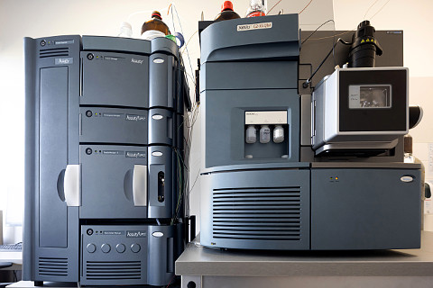
It accurately measures blood serum lipid concentrations using state-of-the-art analytical techniques. The method combines high-performance supercritical fluid chromatography and mass spectrometry to accurately analyse more than 150 lipids that make up the lipid profile. The lipid profiles are then compared using multivariate statistical analysis, from which the test result is determined.
This new testing method is completely non-invasive and poses no risk to the patient. Like a conventional blood test, the LDPC test only requires the collection of venous blood.
Existing diagnostic methods for detecting pancreatic cancer
Pancreatic cancer is so far diagnosed using the following gold standard methods:
EUS (endoscopic ultrasound)
In this method, an endoscopic ultrasound probe is orally inserted. Sound waves are used to take images of the tumour from inside the body, close to the pancreas, increasing the resolution compared to conventional ultrasound. At the same time, targeted tissue samples can be taken to discover the exact type of neoplasm (a biopsy). EUS produces the best results when detecting the early stages of pancreatic cancer.
MR/MRCP (magnetic resonance imaging with magnetic cholangiopancreatography)
This method uses a magnetic field to take detailed images of the body. This kind of examination can provide information about the size of the tumour, the stage of the cancer and can detect abnormal findings.
CT scan (computed tomography)
A CT scan uses X-rays to take cross-sectional images of the body from different angles. The patient may also be given a contrast agent to make it easier to see the organs. This method works well for imaging the pancreas and provides information about the size and location of the tumour and its distribution throughout the body.
Tumour markers
Tumour markers can be used to confirm the presence of a tumour and to monitor the progress of treatment. For example, the concentration of the protein CA 19-9 in the blood can be measured. However, markers for pancreatic cancer, provide limited sensitivity and specificity, especially in the early stages.